
Lab Manual for Human Anatomy and Physiology: A Comprehensive Guide
This comprehensive lab manual provides an in-depth exploration of human anatomy and physiology through structured exercises and interactive activities‚ ensuring a hands-on understanding of the human body’s intricate systems.
Welcome to the Lab Manual for Human Anatomy and Physiology! This guide is designed to enhance your understanding of the human body through hands-on laboratory activities. It provides a structured approach to exploring complex anatomical and physiological concepts‚ making learning engaging and effective. Each section is crafted to align with coursework‚ offering detailed explanations‚ diagrams‚ and practical exercises. Whether you’re a student or an aspiring healthcare professional‚ this manual serves as a valuable resource to master the fundamentals of human anatomy and physiology. Use it to deepen your knowledge and develop essential laboratory skills.
Importance of Anatomy and Physiology in Healthcare Education
Understanding anatomy and physiology is fundamental for healthcare professionals‚ as it forms the basis of diagnosis‚ treatment‚ and patient care. These disciplines provide essential insights into how the human body functions‚ enabling professionals to identify abnormalities and develop effective interventions. A strong foundation in anatomy and physiology also enhances clinical decision-making and communication among healthcare teams. For students pursuing careers in medicine‚ nursing‚ or allied health sciences‚ mastering these subjects is crucial for delivering high-quality‚ patient-centered care. This lab manual is designed to support healthcare education by offering practical exercises that reinforce theoretical knowledge and prepare students for real-world clinical applications.
Lab Safety and Equipment Handling
Lab safety is critical to ensure a secure learning environment. Always handle microscopes with care‚ keeping the objective lens at least 1 cm above the stage. After use‚ store the microscope in its box. Properly clean and maintain equipment to preserve accuracy. Wear lab coats and gloves when handling specimens or chemicals. Follow all safety protocols during experiments to prevent accidents. Dispose of biological waste correctly and label all samples clearly. Familiarize yourself with emergency procedures‚ such as eye wash stations and fire extinguishers. Adhere to these guidelines to create a safe and effective learning atmosphere for all students.
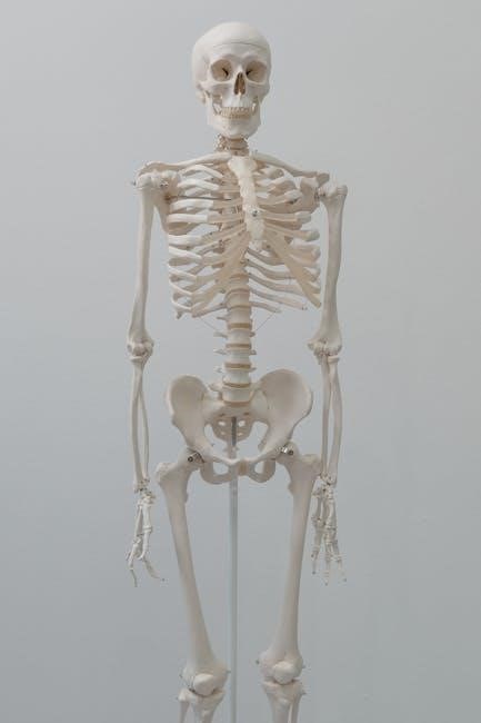
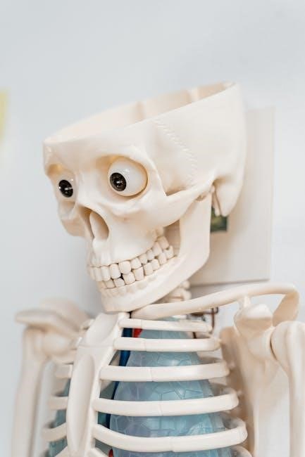
The Skeletal System
The skeletal system provides structural support‚ protects vital organs‚ and facilitates movement through muscles and joints. It also produces blood cells essential for circulation.
Structure and Function of Bones
Bones are rigid‚ calcified tissues forming the skeleton’s framework. Their structure includes a dense outer layer (cortical bone) and a spongy inner layer (trabecular bone). The periosteum covers the surface‚ while the endosteum lines cavities. Bones support the body‚ protect organs‚ and facilitate movement through joints. They also house bone marrow‚ which produces blood cells. The unique combination of collagen and minerals like calcium and phosphorus gives bones strength and flexibility. Bone remodeling continuously replaces old tissue‚ maintaining strength and calcium balance. Understanding bone structure is crucial for studying skeletal health and disorders‚ such as fractures or osteoporosis‚ in anatomy and physiology labs.
Classification of Bones: Axial and Appendicular Skeletons
The human skeleton is divided into the axial and appendicular systems. The axial skeleton includes the skull‚ vertebral column‚ ribcage‚ and sternum‚ primarily protecting vital organs. The appendicular skeleton comprises the upper and lower limbs‚ shoulders‚ and pelvic girdles‚ facilitating movement and support. Bones are further classified by shape: long (e.g.‚ femur)‚ short (e.g.‚ carpals)‚ flat (e.g.‚ skull bones)‚ irregular (e.g.‚ vertebrae)‚ and sesamoid (e.g.‚ patella). This classification aids in understanding their structural and functional roles‚ essential for anatomy and physiology studies. Lab exercises often involve identifying and categorizing bones to reinforce these concepts effectively.
Types of Joints and Their Movements
Joints‚ or articulations‚ are classified based on their ability to move. Synarthroses are immovable joints‚ like skull sutures. Amphiarthroses allow limited movement‚ such as intervertebral discs. Diarthroses are freely moving joints‚ like knees and shoulders. Movements include flexion‚ extension‚ abduction‚ adduction‚ rotation‚ and circumduction. Ball-and-socket joints enable the widest range of motion‚ while hinge joints‚ like elbows‚ allow movement in one plane. Lab exercises involve identifying joint types and their movements‚ using models or palpation‚ to understand their functional roles in the skeletal system. This knowledge is crucial for grasping human mobility and anatomical mechanics.

The Muscular System
The muscular system consists of skeletal‚ smooth‚ and cardiac muscles‚ each performing distinct roles in movement‚ support‚ and maintaining bodily functions; Lab exercises focus on identifying and understanding muscle types‚ their structures‚ and functions through dissection and microscopic analysis‚ providing hands-on insights into their contributions to overall physiology.
Types of Muscles: Skeletal‚ Smooth‚ and Cardiac
The muscular system comprises three distinct types: skeletal‚ smooth‚ and cardiac muscles. Skeletal muscles are voluntary‚ attached to bones‚ and responsible for movement. Smooth muscles are involuntary‚ found in internal organs‚ and regulate functions like digestion. Cardiac muscles are involuntary‚ specific to the heart‚ and ensure rhythmic contractions. Lab exercises involve identifying these muscle types under microscopes‚ exploring their histological structures‚ and understanding their roles in movement‚ organ function‚ and blood circulation. This hands-on approach enhances comprehension of their unique characteristics and contributions to overall physiology.
Structure and Function of Muscles
Muscles are composed of muscle fibers‚ each surrounded by a plasma membrane called the sarcolemma. Inside‚ myofibrils contain actin and myosin filaments that slide to produce contractions. This process‚ regulated by neurotransmitters and ATP‚ enables movement; Muscles function to move bones‚ stabilize joints‚ and facilitate bodily functions like digestion and circulation. Lab exercises involve observing muscle histology under microscopes and performing contraction experiments. Understanding muscle structure and function is crucial for grasping human movement and the impact of muscle disorders. These labs provide hands-on insights into the cellular and physiological mechanisms of muscle activity.
Muscle Actions and Their Roles in Movement
Muscles produce movement through contraction‚ working in groups as agonists‚ antagonists‚ synergists‚ and stabilizers. Agonists initiate movement‚ while antagonists oppose it. Synergists assist‚ and stabilizers maintain joint stability. Lab activities‚ such as muscle palpation and joint movement analysis‚ demonstrate these roles. Understanding muscle actions is vital for analyzing human movement and addressing clinical conditions like injuries or impaired mobility. These exercises help students correlate muscle function with functional anatomy‚ enhancing their ability to apply physiological principles in real-world scenarios. Hands-on exploration of muscle dynamics deepens comprehension of movement mechanics and their significance in maintaining bodily functions and overall physical performance.

The Nervous System
Structure and Function of Neurons
Neurons are specialized cells transmitting nerve impulses through dendrites‚ cell bodies‚ and axons. Lab exercises involve identifying neuronal structures under microscopes and exploring their roles in signaling pathways.
Neurons are specialized cells designed for communication‚ consisting of dendrites‚ a cell body‚ and an axon. Dendrites receive signals‚ while the axon transmits them to other neurons or cells. The cell body contains organelles essential for neuronal function‚ including the nucleus and mitochondria. Lab activities involve studying neuron structures under microscopes and simulating signal transmission. Understanding neuron physiology is crucial for grasping nervous system operations‚ including reflexes and sensory responses.
Types of Nervous Tissue and Their Functions
Nervous tissue consists of two primary types: gray matter and white matter. Gray matter contains neuron cell bodies and is responsible for processing information‚ while white matter is composed of myelinated axons that facilitate rapid signal transmission. Both types work together to enable sensory perception‚ motor responses‚ and complex cognitive functions. Lab exercises often involve histological examinations of nervous tissue samples to identify these structures. Understanding their roles is essential for grasping nervous system physiology and its integration with other body systems.
Reflexes and Nerve Impulses
Reflexes are automatic responses to stimuli‚ involving a sensory neuron‚ the central nervous system‚ and a motor neuron. Lab activities demonstrate reflex actions‚ such as withdrawing a hand from heat‚ showcasing the nervous system’s rapid response mechanism. Nerve impulses‚ or action potentials‚ are electrical and chemical signals that transmit information. They rely on ion exchange across cell membranes and are enhanced by myelin sheaths for faster conduction. Labs often include simulations or histological studies of nerve tissue to visualize these processes. Understanding reflexes and nerve impulses is key to grasping nervous system functionality and its role in controlling bodily functions.

The Circulatory System
The circulatory system transports oxygen‚ nutrients‚ and waste through blood vessels‚ powered by the heart. Lab exercises explore blood composition‚ heart function‚ and vessel roles in maintaining homeostasis.
Components of Blood and Their Functions
Blood is a vital fluid composed of plasma‚ red blood cells (RBCs)‚ white blood cells (WBCs)‚ and platelets. Plasma‚ the liquid portion‚ transports nutrients‚ hormones‚ and waste. RBCs‚ containing hemoglobin‚ carry oxygen throughout the body. WBCs are crucial for immune defense‚ fighting infections and diseases. Platelets play a key role in blood clotting‚ preventing excessive bleeding. Lab exercises involve analyzing blood smears‚ identifying cell types‚ and understanding their roles in maintaining homeostasis. This section provides detailed insights into the structure and function of blood components‚ essential for healthcare education and clinical applications.
Structure and Function of the Heart
The heart is a muscular‚ four-chambered organ divided into the right and left atria and ventricles. The septum separates the chambers‚ while valves ensure blood flows in one direction. The myocardium‚ the thick middle layer‚ contracts to pump blood‚ while the epicardium protects the heart. The heart acts as a dual pump‚ sending deoxygenated blood to the lungs and oxygenated blood to the body. The cardiac cycle includes systole (contraction) and diastole (relaxation). Lab exercises involve dissecting heart models‚ observing blood flow‚ and measuring heart rates to understand its critical role in circulation and oxygen delivery.
Types of Blood Vessels and Their Roles
Blood vessels include arteries‚ veins‚ and capillaries‚ each with distinct roles. Arteries‚ thick-walled and muscular‚ carry oxygen-rich blood away from the heart to tissues. Veins‚ thinner and less muscular‚ return deoxygenated blood to the heart‚ aided by one-way valves. Capillaries‚ tiny and thin-walled‚ facilitate gas and nutrient exchange between blood and tissues. Lab activities involve identifying vessel types through dissection and microscopy‚ exploring their structural adaptations‚ and simulating blood flow dynamics. Understanding these vessels is crucial for grasping circulatory function and oxygen delivery mechanisms in the body.
The Respiratory System
The respiratory system‚ comprising the nose‚ trachea‚ and lungs‚ facilitates breathing and gas exchange. Lab exercises include dissections‚ simulations‚ and measurements to study its functions.
Structure and Function of the Nose‚ Trachea‚ and Lungs
The nose contains nasal passages lined with mucous membranes and cilia‚ filtering‚ warming‚ and humidifying inhaled air. The trachea‚ or windpipe‚ is a flexible tube with cartilaginous rings‚ preventing collapse and leading to bronchi. Lungs are spongy organs divided into lobes‚ facilitating gas exchange via alveoli. Lab exercises include dissections and microscopy to explore these structures‚ while ventilation simulations demonstrate airflow and oxygen-carbon dioxide exchange. Understanding these components is crucial for grasping respiratory physiology and diagnosing disorders.
Process of Ventilation and Gas Exchange
Ventilation involves the inhalation and exhalation of air‚ regulated by diaphragm and intercostal muscle contractions. Inhalation expands lung volume‚ drawing air into alveoli‚ while exhalation expels stale air. Gas exchange occurs in alveoli‚ where oxygen diffuses into blood and carbon dioxide into lungs for exhalation. Lab experiments‚ such as spirometry‚ measure lung capacity and ventilation rates‚ demonstrating respiratory efficiency. Understanding this process is vital for diagnosing respiratory disorders and appreciating the body’s oxygenation mechanisms.
Measurement of Lung Capacity and Volume
Measuring lung capacity and volume involves assessing how much air the lungs can hold and exchange. Techniques like spirometry are used to record tidal‚ inspiratory‚ and expiratory volumes. Vital capacity‚ the maximum air exhaled after full inhalation‚ is crucial for diagnosing respiratory conditions. Lab exercises often include inhaling and exhaling through a spirometer to measure these values. These measurements help understand respiratory efficiency and overall lung health‚ providing insights into conditions like asthma or COPD. Accurate data collection ensures reliable results for clinical and educational purposes.
The Digestive System
The digestive system processes food through ingestion‚ mechanical and chemical breakdown‚ absorption of nutrients‚ and elimination of waste. Key organs include the mouth‚ esophagus‚ stomach‚ and intestines‚ with enzymes playing a crucial role in digestion and nutrient absorption.
Structure and Function of the Mouth‚ Esophagus‚ and Stomach
The mouth initiates digestion with teeth grinding food and saliva adding enzymes like amylase. The esophagus transports food to the stomach via peristalsis. The stomach churns food with gastric juices‚ breaking it into chyme for nutrient absorption in the intestines. Its lining secretes mucus to protect from acid damage‚ ensuring proper digestion and absorption of nutrients for energy and growth.
Role of the Small and Large Intestines in Digestion
The small intestine is the primary site for nutrient absorption‚ facilitated by finger-like projections called villi that increase surface area. Pancreatic enzymes and bile from the liver break down carbohydrates‚ proteins‚ and fats into absorbable molecules. The large intestine absorbs water and electrolytes‚ forming solid waste. It also houses gut flora‚ which aids in vitamin synthesis and fiber digestion. Together‚ the small and large intestines ensure efficient nutrient uptake and waste preparation for elimination‚ maintaining overall digestive health and energy production.
Enzymes and Their Functions in Digestion
Enzymes are biological catalysts that accelerate chemical reactions in digestion‚ breaking down complex nutrients into smaller‚ absorbable molecules. Amylases‚ lipases‚ and proteases target carbohydrates‚ fats‚ and proteins‚ respectively. These enzymes are produced in the mouth‚ pancreas‚ and small intestine. They lower activation energy‚ speeding up digestion. Specificity ensures enzymes act only on target molecules‚ preventing self-digestion. Enzyme deficiencies can lead to malabsorption. The gut microbiome also produces enzymes‚ aiding fiber breakdown. Efficient enzyme function is vital for nutrient absorption‚ energy production‚ and overall health.
The Urinary System
The urinary system‚ comprising kidneys‚ ureters‚ bladder‚ and urethra‚ filters blood‚ removes waste‚ and regulates fluid balance. It produces urine‚ maintaining homeostasis through excretion and electrolyte control.
Structure and Function of the Kidneys
The kidneys are bean-shaped organs located in the lower back‚ each containing millions of nephrons‚ the functional units. The outer cortex and inner medulla facilitate filtration. Blood enters via the renal artery‚ filtered through glomeruli‚ and waste is excreted as urine. The kidneys regulate electrolytes‚ maintain acid-base balance‚ and produce hormones like erythropoietin and renin. Their role in filtering blood and managing fluid balance is vital for homeostasis. Lab exercises often involve dissecting kidney models and observing nephron structures under microscopes to understand their complex functions in waste removal and blood purification.
Process of Urine Formation and Excretion
Urine formation begins with glomerular filtration in the nephrons‚ where blood is filtered to remove waste and excess substances. The filtrate then undergoes tubular reabsorption‚ returning essential nutrients and water to the blood. Tubular secretion adds waste products to the filtrate. The resulting urine flows through the collecting ducts into the renal pelvis and ureters‚ storing in the bladder until excretion via the urethra. This process regulates water‚ electrolytes‚ and pH balance. Lab activities often include simulations or microscopy to visualize these steps‚ enhancing understanding of renal physiology and its critical role in maintaining homeostasis.
Components of Urine and Their Analysis
Urine primarily consists of water (95%)‚ urea‚ creatinine‚ amino acids‚ and ions (e.g.‚ sodium‚ potassium). Other components include glucose‚ ketones‚ proteins‚ and blood cells‚ which may indicate health conditions. Urinalysis involves physical‚ chemical‚ and microscopic examinations; Physical tests assess color‚ clarity‚ and specific gravity. Chemical tests detect pH‚ protein‚ glucose‚ and ketones using dipstick methods. Microscopic analysis identifies cells‚ casts‚ and crystals. Abnormal findings‚ such as blood or excess protein‚ suggest kidney or urinary tract issues. Lab exercises demonstrate these tests‚ aiding in understanding normal and pathological urine profiles and their clinical significance in diagnosing diseases.

The Reproductive System
This section explores the male and female reproductive systems‚ focusing on their structures‚ functions‚ and roles in fertility and development through detailed explanations and practical lab exercises.
Structure and Function of Male and Female Reproductive Organs
The male reproductive system includes the testes‚ epididymis‚ vas deferens‚ and penis‚ with the testes producing sperm and the penis facilitating ejaculation. The female reproductive system comprises the ovaries‚ fallopian tubes‚ uterus‚ and vagina‚ with the ovaries producing eggs and the uterus supporting fetal development. Both systems are essential for gamete production and fertilization. The lab manual provides detailed diagrams and exercises to identify and understand the anatomy of these organs‚ emphasizing their roles in reproduction and sexual health. Interactive activities help students visualize the interplay between structures and their physiological functions in both males and females.
Hormones and Their Roles in Reproduction
Hormones play a vital role in regulating reproductive processes in both males and females. In males‚ testosterone‚ produced by the testes‚ is essential for sperm production and secondary sexual characteristics. In females‚ estrogen and progesterone‚ produced by the ovaries‚ regulate the menstrual cycle‚ pregnancy‚ and lactation. The hypothalamus and pituitary gland control these hormones through gonadotropin-releasing hormone‚ follicle-stimulating hormone‚ and luteinizing hormone. These hormonal interactions ensure proper gamete development‚ fertilization‚ and maintenance of pregnancy. The lab manual includes exercises to explore hormone functions‚ such as endocrine system diagrams and case studies on hormonal imbalances and their effects on reproduction.
Process of Fertilization and Development
Fertilization occurs when a sperm fuses with an oocyte‚ forming a zygote. This process involves the sperm’s acrosome reaction‚ enabling it to penetrate the zona pellucida. Once fused‚ the zygote undergoes cleavage‚ dividing into a blastocyst. The blastocyst differentiates into the embryoblast (future fetus) and trophoblast (placental tissue). Implantation in the uterus follows‚ initiating pregnancy. The lab manual provides detailed diagrams and exercises to explore these stages‚ including microscopy of embryonic development and case studies on fertilization techniques. Understanding these processes is critical for grasping human development and reproductive biology.
Integration of Systems and Advanced Topics
This section explores how body systems interact to maintain homeostasis‚ focusing on advanced lab techniques and clinical applications. It prepares students for real-world healthcare scenarios.
Interrelationships Between Body Systems
The human body operates as an intricate network where systems collaborate to maintain homeostasis. For instance‚ the respiratory and circulatory systems work together to deliver oxygen to cells‚ while the nervous system regulates these processes. Each system’s function is interdependent‚ ensuring overall bodily harmony. Understanding these relationships is crucial for grasping how the body responds to health challenges. The lab manual emphasizes these connections through interactive exercises‚ helping students visualize how systems like the muscular and skeletal frameworks enable movement. This holistic approach fosters a deeper appreciation of the body’s complexity and its ability to adapt to internal and external changes.
Advanced Lab Techniques and Tools
Advanced lab techniques involve cutting-edge tools like 3D printers for anatomical models and electron microscopes for tissue analysis. Molecular biology tools such as PCR enhance genetic studies‚ while robotic aids improve dissection efficiency. These tools enable precise and efficient experimentation‚ allowing for innovative exploration of complex physiological processes in human anatomy and physiology.
Preparation for Clinical Applications
This section focuses on translating anatomical and physiological knowledge into practical clinical skills. Through hands-on exercises‚ students gain proficiency in patient assessment‚ diagnostic techniques‚ and therapeutic interventions. Real-world case studies and simulations bridge the gap between theory and practice‚ enhancing critical thinking and decision-making abilities. Emphasis is placed on understanding medical imaging‚ lab diagnostics‚ and patient monitoring tools. These activities prepare learners for healthcare roles by fostering a deep understanding of how body systems interact in clinical scenarios‚ ensuring they are well-equipped to address patient needs effectively.